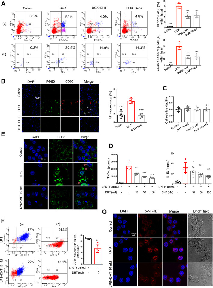
Fig 2.
DHT suppressed the activation of M1 macrophages in mice and in RAW264.7 cells. (A) Flow cytometry assay showed that DHT or Rapamycin reduced accumulation of macrophages and activation of M1 macrophages in mice. In (a), X-axis represents PE-Cy7 anti-CD11b and Y-axis represents APC anti-F4/80; in (b), X-axis represents PE anti-CD86 and Y-axis represents FITC anti-CD206. N ≥ 3 per group. *p < 0.05, **p < 0.01, ***p < 0.001 is significantly different as indicated, for values in the DOX group. b Immunofluorescence assay showed that DHT suppressed the protein expressions of CD86 and F4/80 in mice, scale bar = 20 μm. N = 3 per group. c DHT treatment for 24 h had no cytotoxic effect on RAW264.7 cells at the dosages of 10, 50 and 100 nM. N = 6 per group. d RAW 264.7 cells were stimulated with LPS (1 μg/mL) in the absence or presence of DHT (10, 50 and 100 nM) for 24 h and the releases of TNF-α and IL-1β in cell supernatants were detected by Elisa assay. N ≥ 5 per group. *p < 0.05, ***p < 0.001 is significantly different as indicated, for values in the LPS group. e The protein expression of CD86 was detected by immunofluorescence assay. DHT suppressed the expression of CD86 in RAW264.7 cells, scale bar = 100 μm. N = 12 per group. f DHT reduced activation of M1 macrophages in LPS-stimulated RAW264.7 cells. N ≥ 3 per group. ***p < 0.001 is significantly different as indicated, for values in the LPS group. g DHT suppressed the nuclear localization of p-NF-κB in LPS-stimulated RAW264.7 cells, scale bar = 50 μm. N = 20 per group. All data were presented as means ± SD in triplicate.

