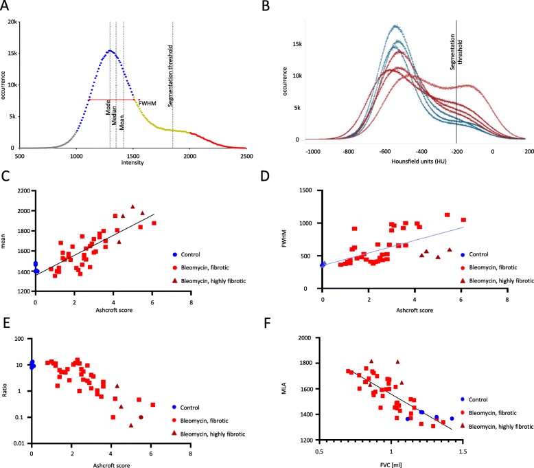
Fig. 3.
μCT data analysis of fibrotic lungs using a novel deep learning approach. (a) An example histogram from μCT data showing indexes used like FWHM and AUC areas (blue and red) for calculation of ratio. b Histogram of μCT data using a novel deep learning approach (control lungs – blue, fibrotic lungs – red). c Correlation of the intensity mean, (d) the FHWM and (e) the AUC ratio of the left lobe (AUC of normal tissue versus highly fibrotic tissue) versus the Ashcroft score. f Correlation of the FVC vs. the intensity mean of the whole lung

