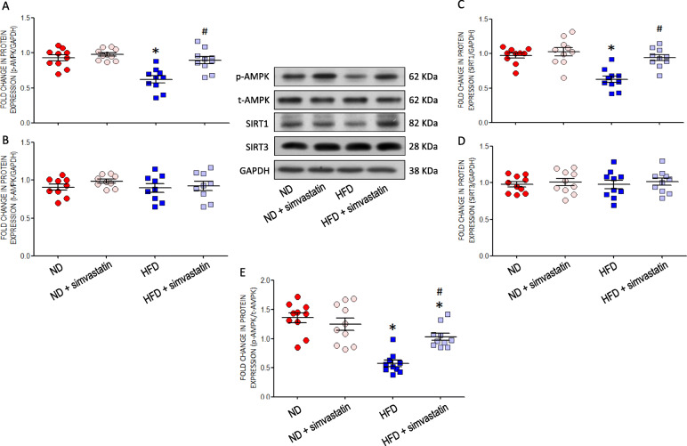
Fig. 4.
AMPK/SIRT signals in RVLM of young offspring exposed to maternal HFD or ND. Representative gels and densitometric analysis of results from Western blot showing changes in protein expression of (a) p-AMPK, (b) t-AMPK, (c) SIRT1 and (d) SIRT3, as well as (e) ratio between p-AMPK/t-AMPK in RVLM of ND or HFD offspring, alone or with additional treatment with simvasatatin (5 mg·kg− 1·day− 1), administered via gastric gavage at age of 8 weeks for 4 weeks. Analysis was performed on tissues collected bilaterally from individual RVLM at age of 12 weeks. Data on protein expression were normalized to the average ND control value, which is set to 1.0, and are presented as mean ± SEM (n = 10 in all groups). *P < 0.05 versus ND group #P < 0.05 versus HFD group in post hoc Newman-Keuls multiple-range test

