
Fig. 3.
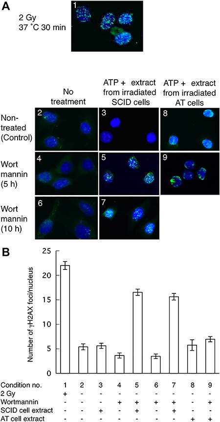
In vitro phosphorylation of histone H2AX. (A) Typical γH2AX foci observed in the nuclei of SCID cells. (B) Numbers of γH2AX foci per nucleus. Panels in (A) and bars in (B) have corresponding numbers designating experimental conditions: 1, SCID cells irradiated and incubated at 37°C for 30 min; 2, no treatment; 3, no treatment with wortmannin and treated with ATP and nuclear extract from irradiated SCID cells; 4, wortmannin-treated (5 h); 5, wortmannin-treated for 5 h and treated with ATP and nuclear extract from irradiated SCID cells; 6, wortmannin-treated (10 h); 7, wortmannin-treated (10 h) and treated with ATP and nuclear extract from irradiated SCID cells; 8, no treatment with wortmannin and treated with ATP and nuclear extract from irradiated AT cells; and 9, wortmannin-treated (5 h) and treated with ATP and nuclear extract from irradiated AT cells. Data are averages of γH2AX foci in 100 cells except in conditions 8 and 9. Conditions 8 and 9 are averages of 25 cells. Error bars indicate the standard error of the number of foci per cell.