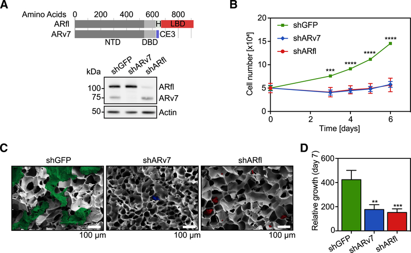
Figure 1. LNCaP95 Cell Growth Is Dependent on both ARfl and ARv7.
(A) Top: schematic of the full-length AR (ARfl) and the AR variant 7 (ARv7), with the N-terminal domain (NTD), DNA-binding domain (DBD), hinge region (H), ligand-binding domain (LBD), and cryptic exon (CE3). Bottom: western blot of shGFP, shARv7, or shARfl LNCaP cells using an N-terminal, pan-AR antibody. Actin signals serve as a loading control.
(B) Proliferation assay of indicated cells grown in 2D culture. Data are the mean of three independent experiments ±SEM.
(C) Representative scanning electron microscopy images of LNCaP95 cells after 7 days of growth in 3D/PEGda cryogels. Scale bar, 100 mm.
(D) Quantification of 3D cell growth data in (C). Data are the mean of four independent experiments relative to day 0 ±SEM.
**p % 0.01; ***p % 0.001; ****p % 0.0001 by Student’s t test. See also Figure S1.

