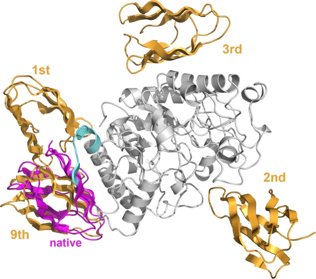
Figure 4.

PDB ID: 1BAG’s receptor domain (gray), 1BAG’s native ligand domain (magenta), four ligand domain models (orange), and the linker region (cyan). This is the only protein in which the permissive structure exists within the top 10 in the prediction based on the interaction amino acid residue score alone. However, the top three structures are far from the native position, and their positions are disjointed. The acceptable model predicted to be 9th. Even for other proteins, predictions with only interaction amino acid residue scores may dislocate the position of the structural model to prevent concentration near the native structure.
