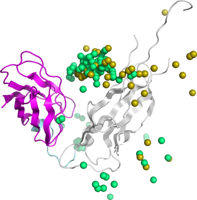
Figure 7.

Native protein structure of 1P5U and center of gravity for top 100 ligands in docking score and PINE. Gray indicates the receptor domain of 1P5U, magenta indicates the ligand domain, cyan indicates the linker region, yellow sphere indicates the center of gravity of the ligand in the structural model generated by initial docking, and green sphere indicates the center of gravity of the ligand in the structural model generated by PINE.
