
Figure 4 – Asymmetry of membrane packing through the endocytic pathway.
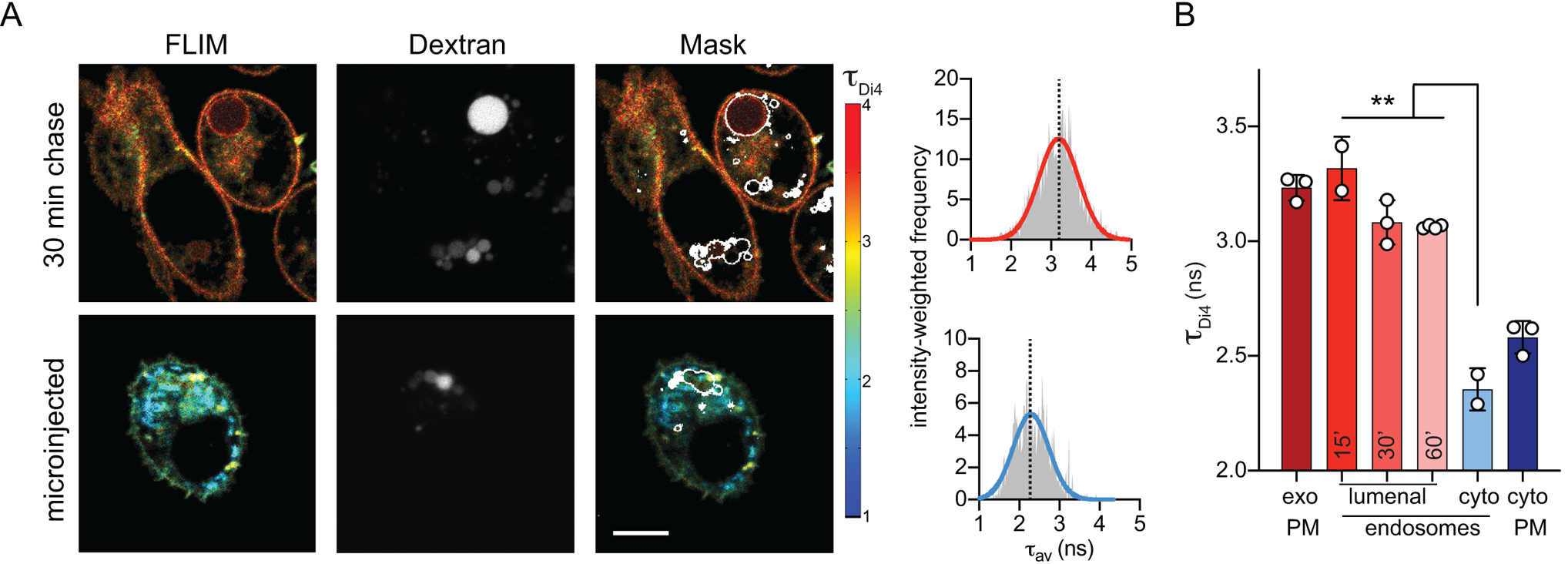
(A) Exemplary FLIM images of Di4 lifetime and dextran fluorescence in RBL cells following 30 min incubation to “chase” stains into endosomes. The accumulation of dextran (middle) is used to create an endosomal membrane mask (right images) to derive intensity-weighted histograms of τDi4 (right). (B) The high lifetime (i.e. lipid packing) of the exoplasmic PM leaflet is maintained in the lumenal leaflets of endosomes, even up to 60 min after endocytosis. Images and quantifications of 3T3 cells are shown in Supplementary Fig. 12. Mean ± SD of >10 individual cells; ***p<0.001 for unpaired t-test. Representative of at least 3 independent experiments.