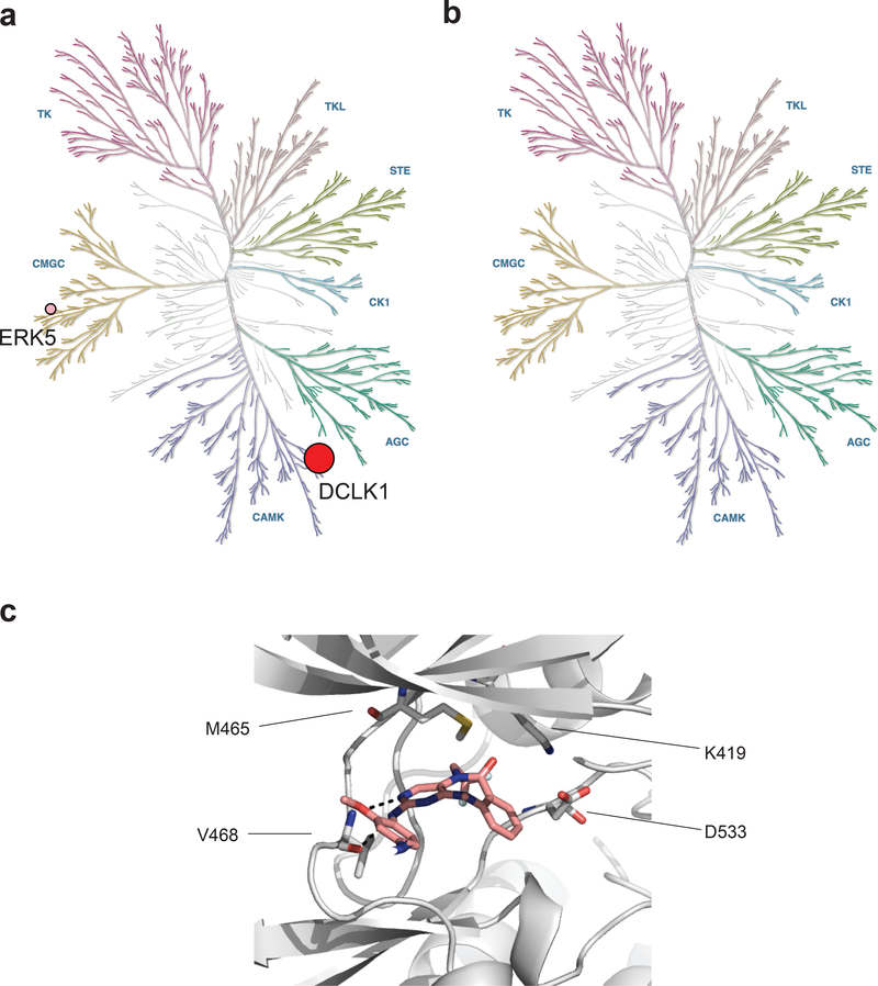
Fig. 2 |. DCLK1-IN-1 engages DCLK1 potently and selectively in cells.
(a-b) KiNativ assay profiling in PATU-8988T cell lysates treated with 2.5 μM DCLK1-IN-1 (a) or DCLK1-NEG (b). The % inhibition represented with circles are 68.5 (DCLK1, red) and 37.9 (ERK5, pink). Data in a-b are presented as the mean of n = 3 technical replicates and associated datasets are provided in Supplementary Dataset 2. (c) Docking model of DCLK1-IN-1 into the X-ray co-crystal structure of the DCLK1 kinase domain (PDB:5JZN). DCLK1 main chain shown grey carbons, DCLK1-IN-1 shown pink carbons. Hydrogen bonds shown as black dashed lines.

