
Figure 1.
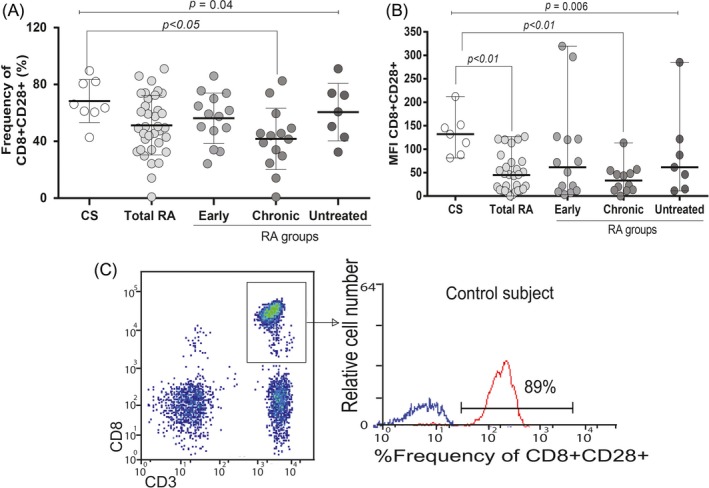
The percentage cell surface expression of CD28 on CD3CD8 + cells of RA patients and CS by flow cytometry. A, Reduced percentage and (B) depicted MFI expression of CD28 on CD3CD8 + cells in chronic RA patients. The data were analyzed by the Kruskal‐Wallis test. C, A representative example is shown, at first lymphocyte populations were gated according to their forward scatter (FS) and side scatters (SS) characteristics. Further, cells from the lymphocyte gate were subgated based on CD3 expression and CD8 co‐expression. Histogram reflects the portion of CD28 expression within the given CD8 + T‐cell populations