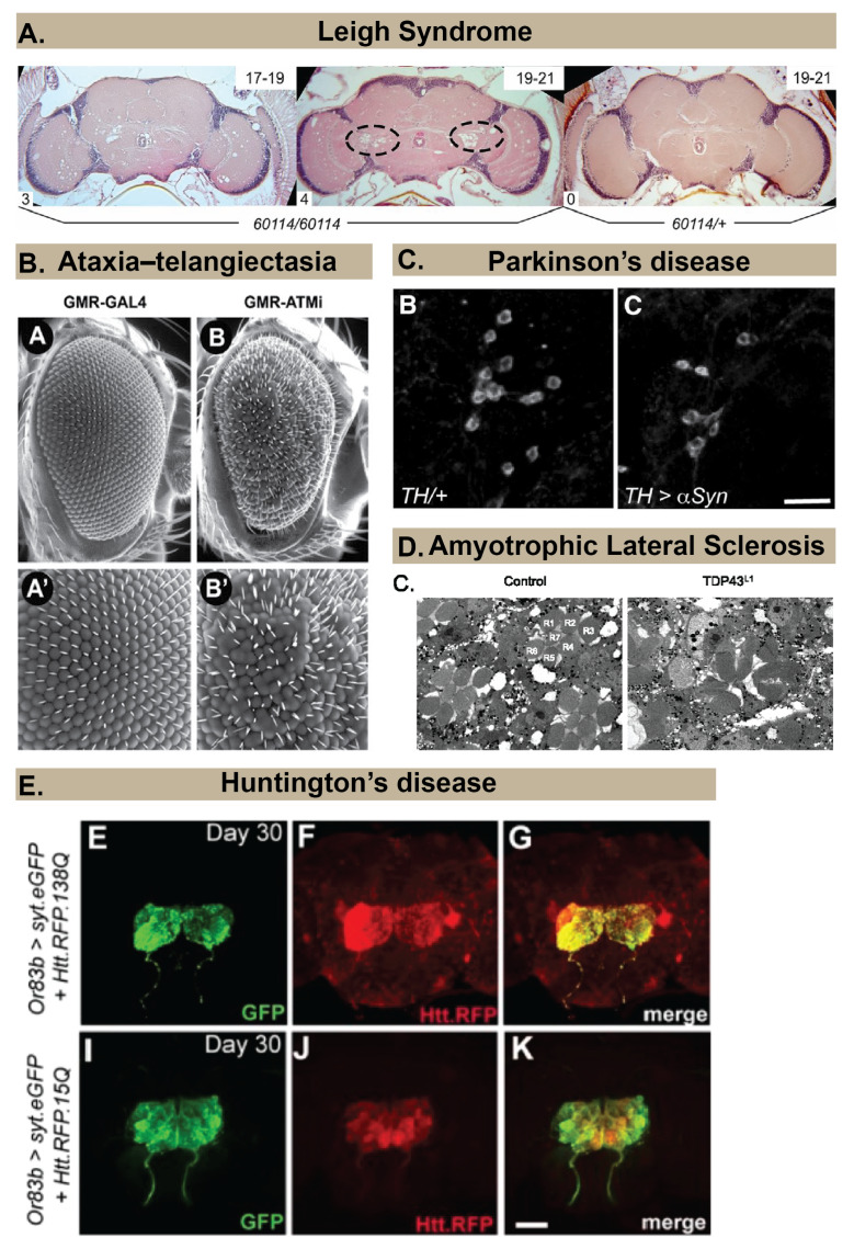
Figure 1.
Examples of approaches to examine neuropathology in Drosophila models of different human neurodegenerative diseases. (A) Spongiform pathology in a Drosophila model of Leigh Syndrome, revealed by histology and hematoxylin and eosin (H&E) staining that shows the appearance of holes in the brain neuropil of 60,114 mutants (ND23 mutants) but not in heterozygous controls (60114/+). Image copyright and permission to use the image were obtained from [21]. (B) Rough eye phenotype (B and B’ for magnified image) observed in a Drosophila model of Ataxia Telangiectasia using scanning electron microscopy. Image copyright and permission to use the image were obtained from [22]. (C) Loss of dopaminergic neurons in a Drosophila model of Parkinson’s Disease is revealed by immunohistochemistry using an anti–Tyrosine Hydroxylase antibody. Image copyright and permission to use the image were obtained from [23]. (D) Neurodegeneration in photoreceptors (labeled R1–R7) of ommatidia in a Drosophila model of Amyotrophic Lateral Sclerosis (right image) is revealed using Transmission Electron Micrographs. Image copyright and permission to use the image were obtained from [24]. (E). Progressive spreading of Red Fluorescent Protein (RFP)-labeled Huntingtin within the brain is revealed by immunohistochemistry in a Drosophila model of Huntington’s Disease. Image copyright and permission to use the image were obtained from [25].

