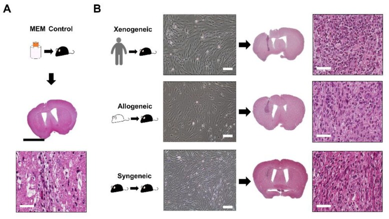
Figure 2.
Transplantation of MSCs in the mouse caudate putamen confirmed via H&E staining. (A) The injection site was identified following MEM injection. CD45-positive leukocytes were barely observed at the injection site. (B) MSCs from all three groups (xenogeneic, allogeneic, and syngeneic) shared typical fibroblastic and proliferative profiles in vitro. Images were taken when cells reached 80%–90% confluency. H&E staining of mice brain tissues transplanted with MSCs from three different sources (xenogeneic, allogeneic, and syngeneic). The injection tract was clearly visible in the caudate putamen of mice (intense dark purple). Scale bars = whole brain: 2 mm, H&E (magnified): 50 µm, and in vitro cell images: 100 µm.

