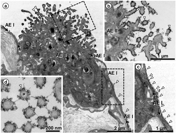
Figure 4.
Human lung. Visualization of the glycocalyx on the surface of alveolar epithelial type I (AEI) and type II (AEII) cells. (a) Overview showing one AEII cell and neighboring thin AEI cell extensions, the lineage of the latter highlighted by arrows. The black lining and dots on the cell surfaces (arrowheads in (a)) depict the glycocalyx, marked by colloidal thorium dioxide. The boxed areas are shown in (b,c) at higher magnification. Note heavily stained microvilli (arrowheads in (b)) and staining at the apical cell membrane of AEI cell (arrowheads in (c)). (d) Profiles of cross-sectioned microvilli (one of them marked by asterisk) of an AEII cell. The glycocalyx (arrowhead in (d)) surrounds the microvilli and also appears as threads between them.

