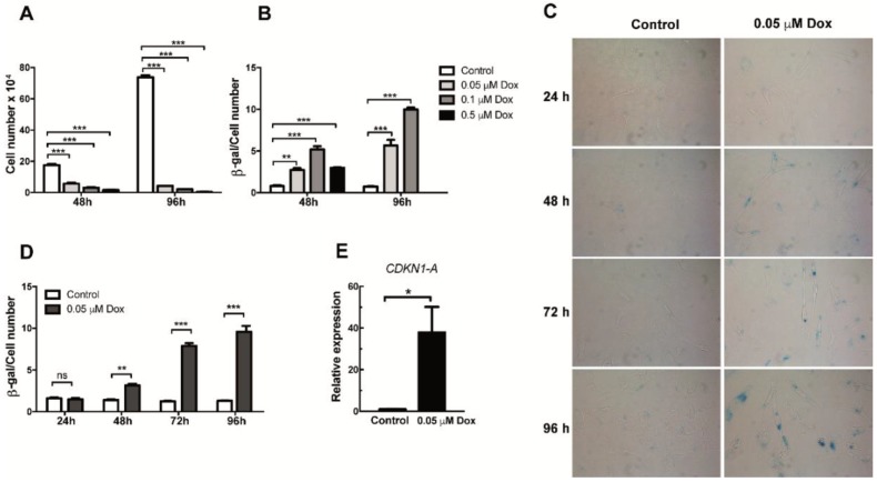
Figure 1.
Analysis of proliferation and senescence in doxorubicin-treated HMEC-1 cells. (A) Number of HMEC-1 cells treated with three different concentrations of doxorubicin for 48 and 96 h. (B) Senescence-associated (SA)-β Galactosidase (SA-β-Gal) activity in doxorubicin (Dox)- and vehicle-treated (control) HMEC-1. Quantification was based on color intensity corrected by the number of cells. (C) Representative images of SA-β-Gal staining in HMEC-1 cells following treatment with 0.05 μM of doxorubicin for 24, 48, 72 and 96 h. (D) Quantification of SA-β-Gal activity in HMEC-1 cell treated with 0.05 μM of doxorubicin for 24, 48, 72 and 96 h. (E) Expression analysis of CDKN1 (encoding p21CIP1/KIP1) RNA levels in cells treated with 0.05 μM of doxorubicin. Error bars indicate mean ± SD of n = 3 (NS = no significant; * p < 0.05; ** p < 0.01; *** p < 0.001; t-student test).

