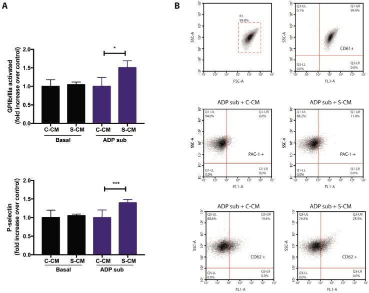
Figure 4.
Effects of media conditioned by senescent cells on platelet activation. (A) The presence of activated GPIIb/IIIa and P-selectin on the surface of platelets, previously exposed to media conditioned by senescent and non-senescent HMEC-1 cells, was determined by flow cytometric analyses. Platelets under basal and sub-aggregating (ADP sub) conditions were tested. (B) Representative dot plots for the detection of CD61 (platelets; top), PAC-1 (activated GPIIb/IIIa; middle) and CD62 (P-selectin; bottom) on human platelets. ADP sub: 1.3–2.0 μM ADP; C-CM: Control conditioned medium; S-CM: Senescent conditioned medium; SSC: side scatter; FSC: forward scatter. The graph depicts the mean ± SD of n = 4 (PAC-1) and n = 7 (CD62) experiments. * p < 0.05 and *** p < 0.001 analyzed by Student’s t-test.

