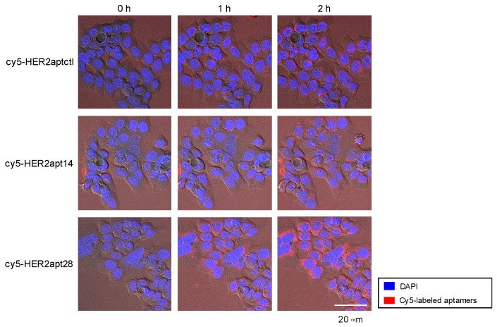
Figure 3.
Confocal microscopic imaging of cy5-HER2apts internalization. NCI-N87 cells (3 × 10⁵) were treated with Cy5-labeled aptamers (cy5-HER2aptctl, cy5-HER2apt14, and cy5-HER2apt28) and DAPI, and incubated at 37 °C for 2 h. The representative are merged images of DIC, DAPI, and Cy5. Internalization of aptamers was visualized using an LSM 780 confocal microscope. Representative fluorescent images were obtained at 0, 1, and 2 h post treatment with cy5-HER2apts. Original magnification, 40×; Scale bar, 20 μm.

