Figure 5.
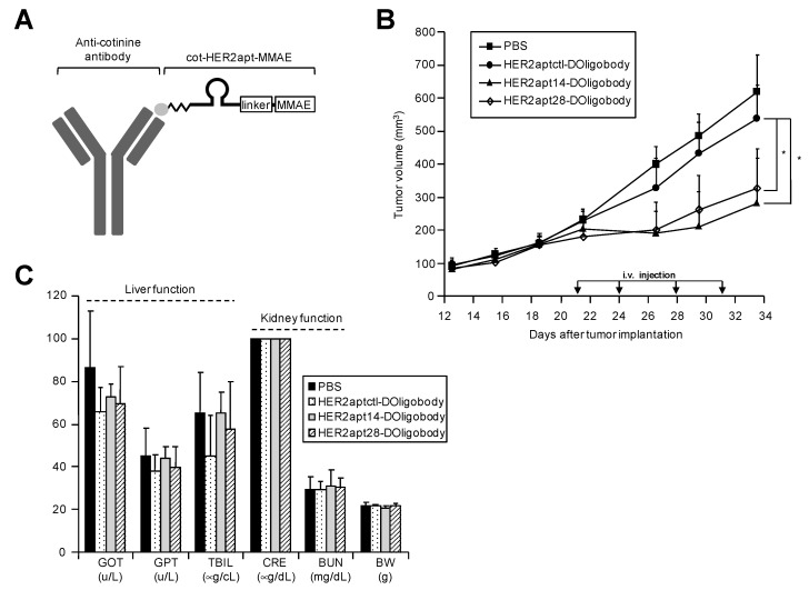
Anti-tumor activity of systemically administered HER2 DOligobodies in a mouse xenograft model. (A) HER2 DOligobody schematic representation. The DOligobody consists of the four elements: the cotinine (cot)-body, cot-linker, aptamer and monomethyl auristatin E (MMAE). (B) NCI-N87 cells (1 × 10⁷) were subcutaneously injected into the flank region of BALB/c nude mice. When the tumors reached 200 mm3, the mice (n = 10 per group) were intravenously injected with PBS (■), control HER2-DOligobody (●), HER2apt14-DOligobody (▲), or HER2apt28-DOligobody (◇) (1.27 mg/kg cot-HER2apt-MMAEs pre-incubated with 10 mg/kg cot-body). Tumor volumes were monitored for 34 d. Data are shown as the mean ± standard error of the mean (SEM); * p < 0.05 compared with the control HER2-DOligobody group, Student’s t-test. (C) In vivo toxicity reflects changes in body weight of the mice and serum concentrations of GOT, GPT, BUN, CRE and TBIL measured 36 d after tumor implantation. All the data represent the means ± SEM from three independent experiments. GOT, glutamic oxaloacetic transaminase; GPT, glutamic pyruvic transaminase; TBIL, total bilirubin; CRE, creatinine; BUN, blood urea nitrogen; BW, body weight.

