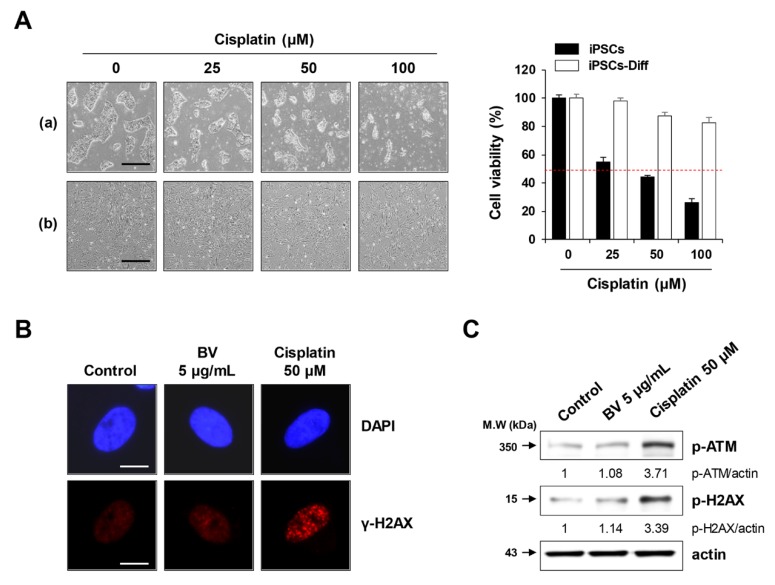
Figure 6.
Effects of BV on the DNA damage in iPSCs-Diff. (A) iPSCs and iPSCs-Diff were seeded in 12-well culture plates and incubated in the presence of cisplatin at 25, 50, and 100 μM for 24 h. Cells were photographed under an inverted microscope and relative cell viability compared with cisplatin-untreated control cells was determined. Data are expressed as means ± SD from triplicate samples. Scale bar = 100 µm. (B) iPSCs-Diff grown on confocal dishes were treated with 5 μg/mL BV or 50 μM cisplatin for 24 h. Cells were stained with anti-p-H2AX antibody followed by Alexa 594-conjugated anti-rabbit antibody. After counterstaining nuclei with DAPI, γ-H2AX foci were observed under a fluorescence microscope. Scale bar = 20 µm. (C) BV- or cisplatin-treated iPSCs-Diff were analyzed for the protein levels of p-ATM and p-H2AX by Western blotting. Data are representative of two independent experiments.

