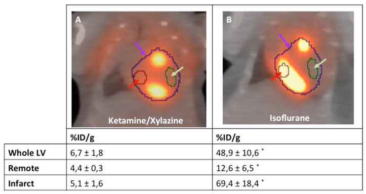
Figure 4.
Representative examples of the analysis strategy underlying the protocol for both mice anesthetized with isoflurane (A) and ketamine/xylazine (B) 5 days after MI induction (n = 4 per group). The “entire left ventricle” VOI reflects the global FDG uptake of the LV (purple arrow). The “remote” VOI was positioned in the inferobasal wall and reflects viable myocardium (red arrow). The “infarct” VOI reflects infarct tissue and contains almost no cardiomyocytes (green arrow). *: p < 0.05 compared to animals anesthetized with ketamine/xylazine. Values are presented as mean ± SD. Values are presented as mean ± SD. p-values were calculated using the student t-test.

