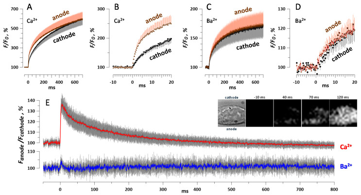
Figure 8.
Polarity effect in the permeabilization of HEK cells by a single 600-ns pulse at 10 kV/cm. Cells were loaded with Calbryte dye and incubated in the physiological solution with 2 mM Ca2+ (A,B) or 2 mM Ba2+ (C,D). The acquisition rate was 3048 frames/s and the pulse was delivered at zero time point. Panels B and D show the same traces as A and C, respectively, at a higher time resolution. Panel E shows the difference in the fluorescence measured at the anode- and cathode-facing poles of each individual cell, averaged for all cells in the group. The inset illustrates preferential anodic entry of Ca2+ in a representative cell. The first image taken in the bright field shows the cell boundaries and the directions to the cathode and anode electrodes relative to the cell. The next images show the Calbryte fluorescence signal at indicated times before and after nsPEF. All images are 17 × 17 µm. Mean +/− s.e., for nine experiments with Ba2+ and 13 with Ca2+; error bars are plotted in one direction only, except for panel E. The difference between Ca2+ readings from the opposite poles (E) is significant at p < 0.01 (one-sample t-test).

