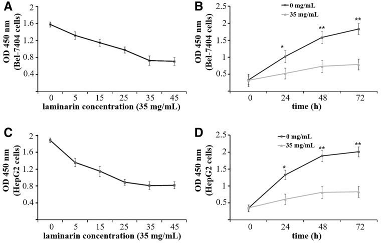
FIG. 1.
The viability (OD at 450 nm) of Bel-7404 and HepG2 cells detected by WST-8 cell proliferation assay. (A) Bel-7404 cells treated with laminarin at different concentrations for 48 h. (B) Bel-7404 cells treated with 35 mg/mL laminarin for different times. (C) HepG2 cells treated with laminarin at different concentrations for 48 h. (D) HepG2 cells treated with 35 mg/mL laminarin for different times. * and **Represent significantly different at p < 0.05 and p < 0.01 when compared with 35 mg/mL laminarin, respectively.

