
Fig. 1.
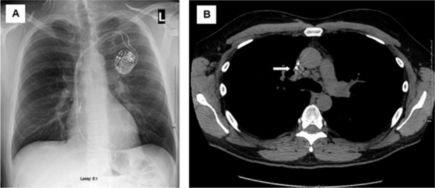
Initial chest imaging in 47-year old male (subject 17) subsequently diagnosed with cardiac sarcoidosis. A. Chest x-ray showing no significant abnormalities. B. Computed tomography of the chest showing mediastinal lymphadenopathy (white arrow)