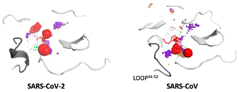
Figure 4.
Localisation of the local hot-spots identified in the binding site cavities in SARS-CoV-2 and SARS-CoV main proteases. Hot-spots of individual cosolvents are represented by spheres, and their size reflects the hot-spot density. The colour coding is as follows: purple—urea, green—dimethylsulfoxide, yellow—methanol, orange—acetonitrile, pink—phenol, red—benzene. The active site residues are shown as red sticks, and the proteins’ structures are shown in cartoon representation; loop 44–52 is grey. The proteins’ structures come from the MD simulation snapshots (first frame of the production stage).

