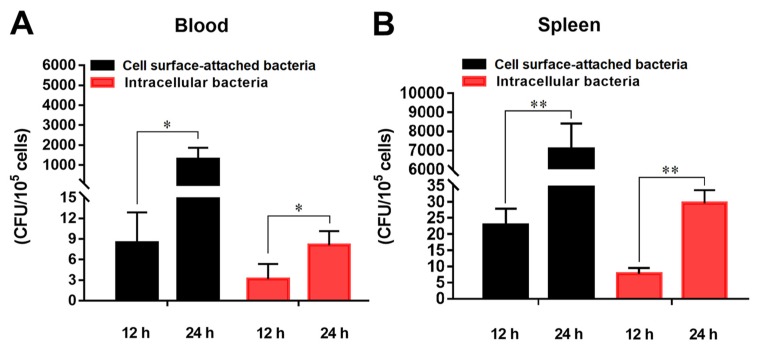
Figure 2.
In vivo infection of Edwardsiella tarda in flounder blood and spleen erythrocytes. Flounder were infected with or without (control) E. tarda for 12 h and 24 h, and erythrocytes were collected from blood (A) and spleen (B). The cell surface-attached and intracellular E. tarda were determined and shown as Colony Forming Unit (CFU). Data are presented as means ± SEM of three independent experiments. *, p < 0.05; **, p < 0.01.

