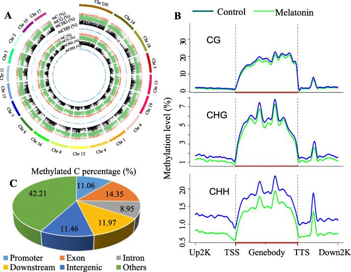
Fig. 2.
Methylation levels of different chromosomes and genomic regions in ‘Merlot’ berries. a The outermost bold lines indicate different chromosomes and their lengths at a 50 kilobase resolution. Red, green, black and blue peak shape diagrams indicate the methylation levels of mC, mCG, mCHG and mCHH, respectively, at different chromosome sites, and the peak height indicates the methylation level. The second to fifth circles are from control berries, and the sixth to ninth circles are from melatonin-treated berries. b Changes in the levels of CG, CHG and CHH methylation in melatonin-treated berries compared to the control. TSS, transcription start site. TTS, transcription termination site. Up2K and Down2K represent the 2000 bp upstream of the TTS and downstream of the TTS, respectively. c Percentages of differentially methylated cytosines in different genomic regions

