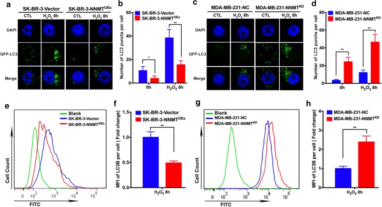
Fig. 3.
NNMT decreased LC3 puncta formation induced by H2O2. a, c The LC3 puncta in the two cell models after H2O2 treatment were imaged by confocal microscopy. A representative result from three independent experiments. b, d The quantification results of (a) and (c), respectively (**p < 0.01 and ***p < 0.001). e, g The fluorescence intensity of LC3B in the two cell models after H2O2 treatment was assessed by flow cytometry. A representative result from three independent experiments. f, h The quantification results of (e) and (g), respectively (**p < 0.01 and ****p < 0.0001)

