
Figure 5.


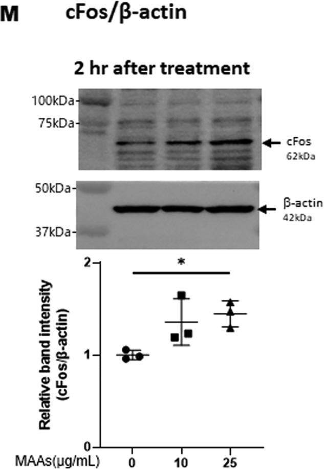
Effects of MAAs on intracellular signaling cascades of p38 (A), ERK (B), JNK (C), ATF2 (D), MSK1(Thr-581/Ser-360/Ser-376) (E–G), c-Jun (H), NF-κB (Ser-276/Ser-536) (I and J), IκB (K), CREB (L), and c-Fos (M) in HDFs. HDFs were incubated with MAAs or with IL-1α at the indicated concentrations and were harvested at 15 and 30 min or 2 h post-treatment and then immunoblotted with antibodies to β-actin and to phosphorylated or nonphosphorylated signaling factors. K: β-actin was used as a loading control because of undetectable levels of nonphosphorylated IκB protein in IL-1α–treated cells. Representative immunoblots from three independent experiments are shown. Data represent means ± S.D., n = 3; *, p < 0.05; **, p < 0.01 versus control (0 μg/ml).