Figure 3.
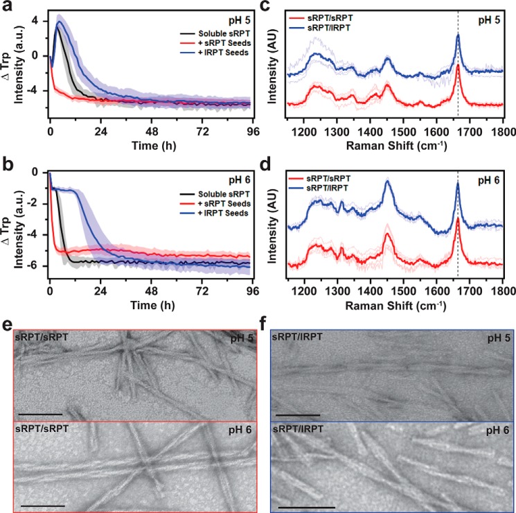
Cross-seeding reactions of sRPT by preformed lRPT fibrils. a and b, aggregation of 30 μm sRPT in the absence (black, unseeded) and in the presence of 3 μm preformed sRPT (red, self-seeding) or lRPT (blue, cross-seeding) fibrils at pH 5 and 6. Reactions were monitored under constant linear shaking (6 mm) at 37 °C. Solid line and shaded area represent the mean and S.D., respectively, from ≥5 replicates. See Fig. S7 for additional dataset. c and d, self- (sRPT/sRPT, red) and cross-seeded (sRPT/lRPT, blue) fibrils, formed at pH 5 and 6, were analyzed by Raman spectroscopy. Spectra were collected at multiple spatial locations (thin lines), which were then averaged (bold line). The vertical dashed line represents 1665 cm−1, to which data were normalized. Cross-seeded spectra are offset for clarity. e and f, representative TEM images of sRPT/sRPT and sRPT/lRPT fibrils. Scale bars represent 100 nm.

