Figure 7.
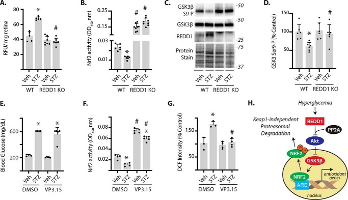
REDD1-dependent GSK3 activation contributes to diabetes-induced oxidative stress and suppression of Nrf2 activity in the retina. Diabetes was induced in WT and REDD1 knockout (KO) mice by administration of STZ. All analyses were performed 4 weeks after mice were administered STZ or a vehicle control (Veh). A, ROS was quantified in the supernatants from whole retinal lysates using dihydroethidium. B, Nrf2 activity was measured in the nuclear fraction obtained from retinal lysates by DNA-binding ELISA. C, GSK3β phosphorylation, as well as expression of GSK3β and REDD1 in retinal lysates were evaluated by Western blotting. Protein loading was evaluated by reversible protein stain. Protein molecular mass (in kilodaltons) is indicated at the right of blots. D, GSK3β phosphorylation in C was quantified. E–G, mice were treated daily with either VP3.15 or a vehicle control (DMSO) during the 4th week of diabetes. E, blood glucose concentrations were evaluated prior to retinal extraction. F, Nrf2 activity was measured as described in B. G, ROS was quantified in the supernatants from whole retinal lysates using DCF. Values are mean ± S.D. *, p < 0.05 versus Veh. #, p < 0.05 versus WT or DMSO. G, working model for the suppressive effect of REDD1 on Nrf2 stability.

