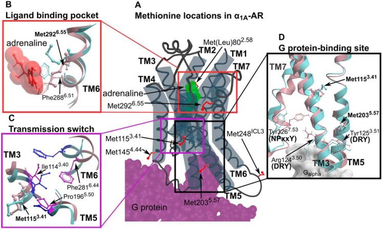
Figure 1.
Methionine residues in α1A-AR. A, the location of six methionines on a cartoon representation of α1A-AR. Methionine side chains are highlighted as red sticks. Bound adrenaline and G protein are colored in green and purple, respectively. B–D, homology models of α1A-AR-A4 in the inactive state (blue; modeled on the X-ray crystal structure of inactive β2-AR, PDB ID 5JQH) and active state (pink; modeled on the X-ray crystal structure of active β2-AR, PDB ID 3SN6) are superimposed showing inferred conformational changes that occur in the ligand-binding pocket (B), transmission switch (C), and G protein-binding site (D).

