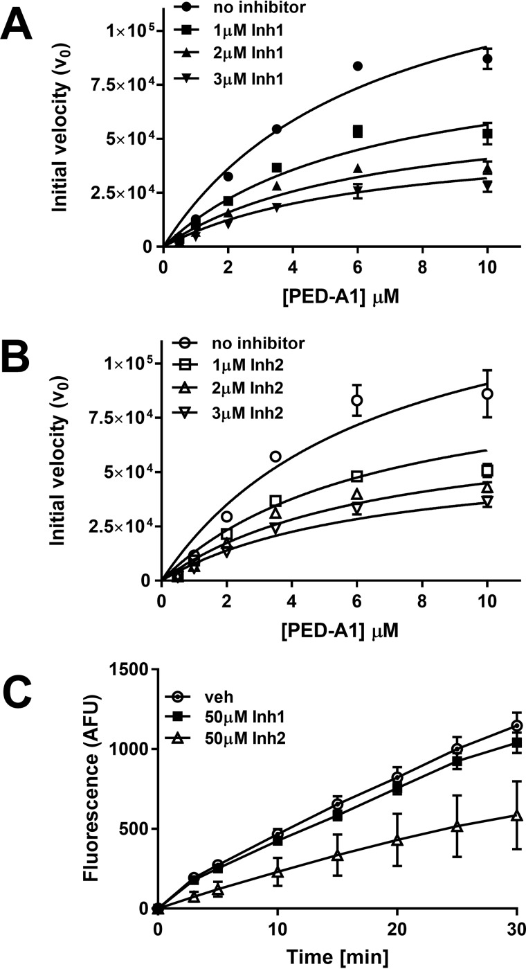
Figure 4.
Mechanism of inhibition by Inh 1 and Inh 2. A, recombinant mouse Nape-pld was incubated with 0–3 μm Inh1, then 0–10 μm PED-A1 was added, and the initial velocity of PED-A1 hydrolysis was determined for each (means ± S.E., n = 3). B, recombinant Nape-pld was incubated with 0–3 μm Inh 2, then 0–10 μm PED-A1 was added, and the initial velocity of PED-A1 hydrolysis was determined for each (means ± S.E., n = 3). C, a rapid dilution assay for Inh 1 and Inh 2. Recombinant Nape-pld was incubated with DMSO (veh) or 50 μm Inh 1 or Inh 2 for 1 h, then samples were diluted 100-fold with buffer immediately prior to addition of 4 μm PED-A1, and the resulting fluorescence was measured as arbitrary fluorescence units (AFU) (means ± S.E., n = 9).

