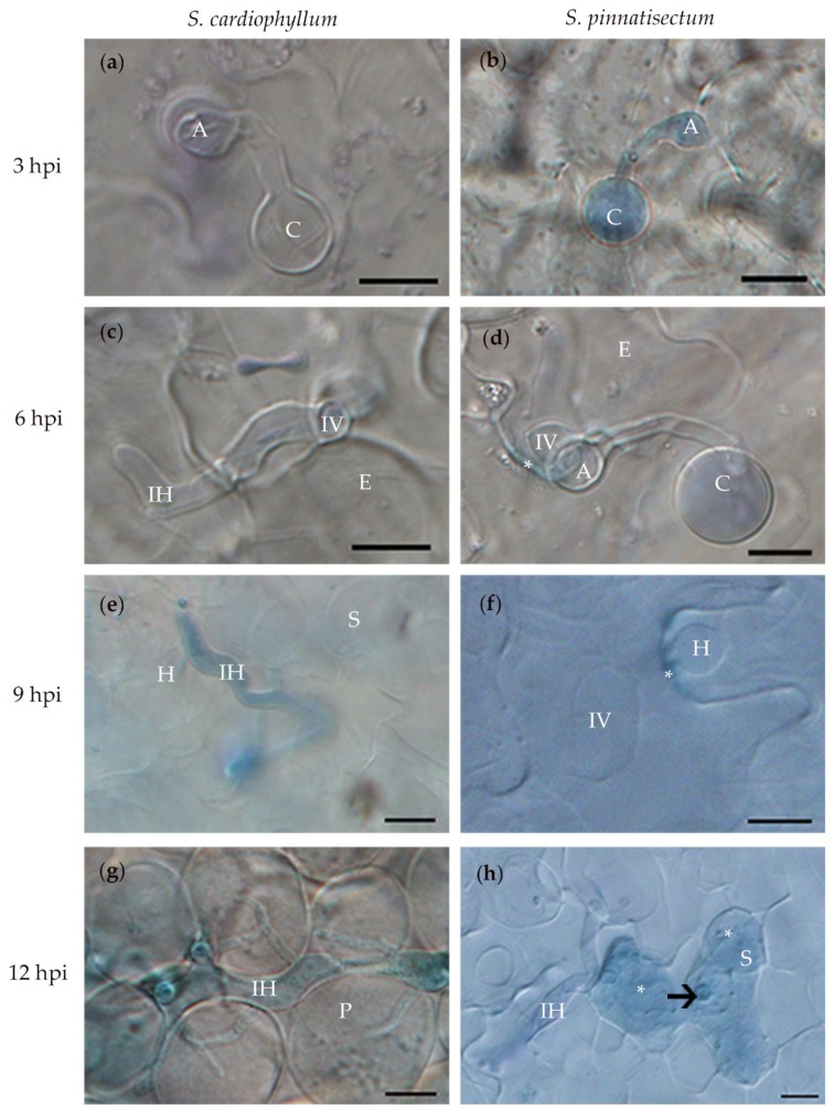
Figure 2.
Microscopic characterization of Solanum pinnatisectum and Solanum cardiophyllum interacted with Phytophthora infestans at different times post-inoculation. (a) and (b), the germinated cysts with appressorium at the tip on the leaf surface of S. cardiophyllum and S. pinnatisectum 3 h post-inoculation (hpi), respectively. (c) and (d), the pathogen penetrated into the epidermal cell of S. cardiophyllum and S. pinnatisectum 6 hpi, * in (d) indicates cell wall deposition at the infection site of epidermal cell on S. pinnatisectum. (e) Intercellular hyphae with haustoria invade spongy cells of S. cardiophyllum 9 hpi. (f) The pathogen extends underneath the cuticle as the infection vessel and forms haustoria in the epidermal cells of S. pinnatisectum 9 hpi. (g) Intercellular hyphae expand with multiple branches in the palisade cells of S. cardiophyllum 12 hpi. (h) Penetration of intercellular hyphae is stopped by the necrosis of mesophyll cells on S. pinnatisectum 12 hpi; the haustorium is pointed by the arrow; and the deeply blue-stained tissues indicate cell necrosis (indicate as *). A: appressorium, C: cyst, E: epidermal cell, H: haustorium, IH: intercellular hyphae, IV: infection vessel, P: palisade cell, S: sponge cell. Bar = 10 μm.

