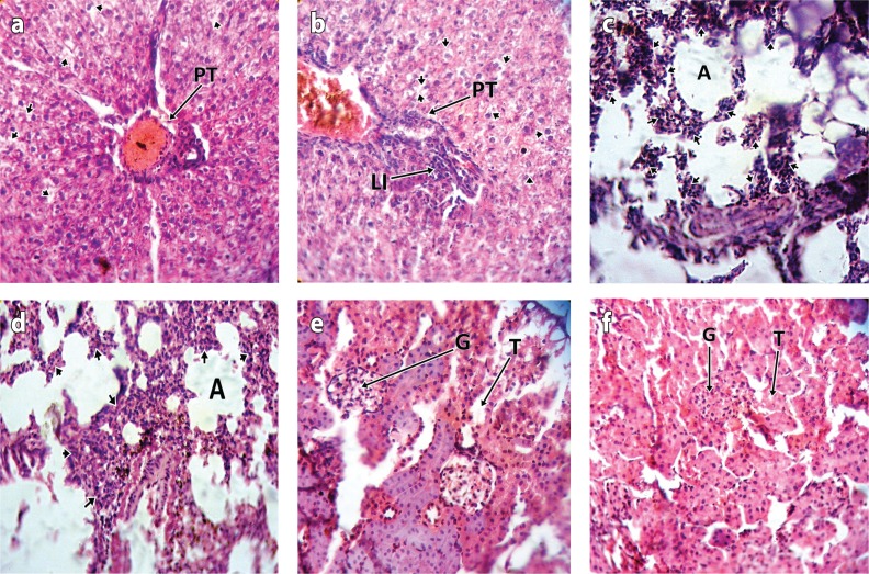
Figure 1.
a: Photomicrograph of the liver of test animal treated with cypermethrin for 2 weeks showing mild steatosis of hepatocytes (short arrows) around portal tract (PT) (H & E stain ×400). b: Photomicrograph of the liver of test animal treated with dichlorvos for 2 weeks showing moderate steatosis of hepatocytes (short arrows) and mild lymphocytic infiltration (LI) of portal tract (PT) (H & E stain ×400). c: Photomicrograph of the lung of test animal treated with cypermethrin for 2 weeks showing moderate lymphocytic infiltration (short arrows) of the interstitium (H & E stain ×400). d: Photomicrograph of the lung of test animal treated with dichlorvos for 2 weeks showing moderate lymphocytic infiltration (short arrows) of the interstitium (H & E stain ×400). e: Photomicrograph of the kidney of test animal treated with cypermethrin for 2 weeks showing unremarkable glomeruli (G) and renal tubules (T) (H & E stain ×400). f: Photomicrograph of the kidney of test animal treated with dichlorvos for 2 weeks showing unremarkable glomeruli (G) and renal tubules (T) (H & E stain ×400).

