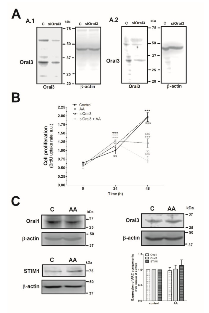
Figure 4.
Combination of AA and siOrai3 reduces MDA-MB-231 cell proliferation. (A) MDA-MB-231 cells were transfected with the siRNA A (control) or siOrai3 for 48 h (A.1) or 96 h (A.2), respectively. WB using the anti-Orai3 antibody was done as described in the Materials and Methods, and following, reprobing of the membranes with an β-actin-antibody was conducted for protein loading control. (B) MDA-MB-231 cells were transfected with the siRNA A (control and AA) or siOrai3 for 48 h. Then, an equal number of cells were shed in a 96-well plate and were allowed to proliferate in the absence or presence of AA (8 μM). At the indicated time points (0, 24, and 48 h), cells were incubated with BrdU for 2 h. The histogram represents the mean ± S.E.M. (standard error of the mean) of BrdU uptake of eight independent experiments. * p < 0.05, ** p < 0.01, *** p < 0.001 respect the BrdU values found in the control at time 0. $$$ p < 0.001 with respect to the BrdU values found in the control at each given time point. ∞ p < 0.05 with respect to the BrdU values found in cells non-genetically modified but treated with AA. We used here the ANOVA and Dunnett’s post-test. (C) MDA-MB-231 cells were cultured for 48 h in the presence or absence of AA (8 μM), and cells were subsequently lysed. Upon protein normalization, WB using the anti-STIM1, anti-Orai1, and anti-Orai3 antibodies were performed as described in the Materials and Methods. Bar graph represents the fold increase ± S.E.M. of the protein expression with respect to the control cells non-treated with AA, which did not report statistical significance upon analyzing with ANOVA and Tukey’s post-test.

