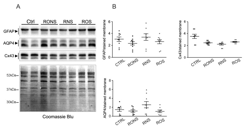
Figure 4.
GFAP, AQP4 and Cx43 immunoblotting and quantification in PALM-incubated astrocytes. (A) Representative Western blot analysis of GFAP, AQP4 and Cx43 expression in astroglial cultures exposed to untreated medium (Ctrl), to P.Air (RNS), to P.O2 (ROS) and to P.N2 (RONS). Protein samples were collected 24 h after 2 h-PALM incubation. (B) Summary of the densitometric analysis of GFAP, AQP4 and Cx43 corresponding signals normalised to the Coomassie blue-stained membrane. No significant differences by One-way Anova test were found between PALM-treated and Ctrl astrocytes.

