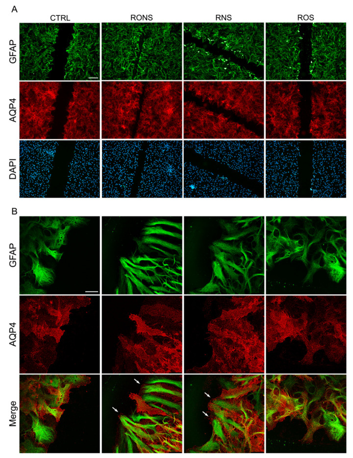
Figure 5.
GFAP and AQP4 protein localisation in astrocyte cultures after PALM incubation. Immunostaining at 6 h after scratch for AQP4 (red) and GFAP (green) in PALM-exposed and control astrocytes. Untreated medium (Ctrl), P.N2 (RONS), P.Air (RNS) and P.O2 (ROS) PALM. (A) Fluorescence signal for AQP4 and GFAP in migrating cells at the wound. Nuclear staining with DAPI is also shown. (Scale bar: 100 μm). (B) Confocal images at the leading edge of wounded astrocytes as in (A). Merged images (Merge) of the two proteins’ localisation are reported (Scale bar: 25 μm). White arrows in RONS and RNS indicate enhanced foot processes at the front end of the scratch.

