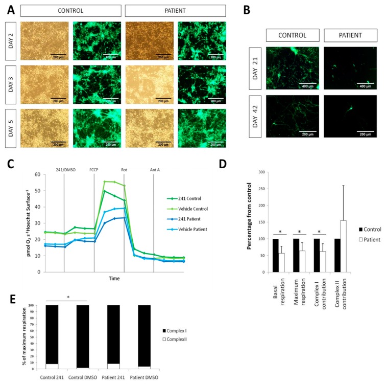
Figure 4.
Respiratory defect and neurodegeneration of patient iPSC-derived neurons. (A) iN generation from iPSCs using lentiviral vectors for NgN2, rtTA and GFP showing no alterations in derivation of iNs from the patient. (B) iNs co-cultured with mouse astrocytes showing a marked neurodegeneration in the patient in comparison with the control both at days 21 and 42. (C) Oxygen consumption plots of the different treatments (Control/Patient and NV241/DMSO). (D) Quantification of oxygen consumption measured in a Seahorse XFe96 Analyzer. All data are normalized with the control. (E) Quantification of the contributions of complexes I and II to the maximum respiration, in percentages. (* p-value < 0.05)

