Figure 1.
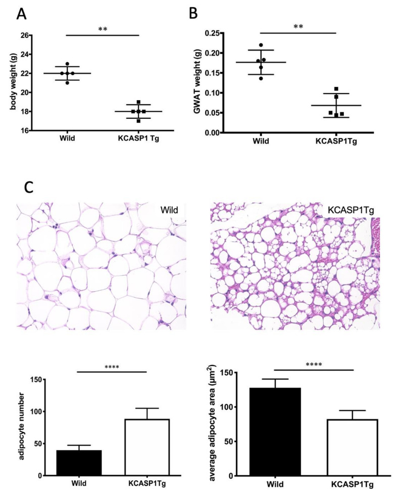
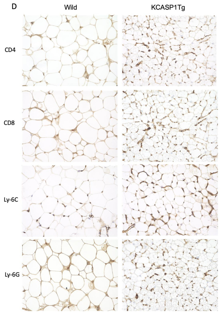
KCASP1Tg mice showed low gonadal white adipose tissue (GWAT) weights and degenerated adipose tissue (AT). (A) The whole body and (B) GWAT weights were decreased in KCASP1Tg compared to wild littermate mice at 10 weeks of age. (C) Representative photomicrographs of the hematoxylin and eosin (HE) (×400) staining of the GWAT of wild littermate and KCASP1Tg mice at 10 weeks of age. Histological sections were 3.5 µm thick. Adipocyte number and adipocyte area were measured by using ImageJ and Adiposoft (three parts of each slide, n = 3 in each group). Adipocytes were significantly smaller and larger in number in KCASP1Tg. (D) Immunohistochemical staining (×400) was performed to characterize stromal cells. Lymphocytes, monocytes and neutrophils were slightly higher in KCASP1Tg but not significantly so. ** p < 0.01, **** p < 0.0001 versus control.


