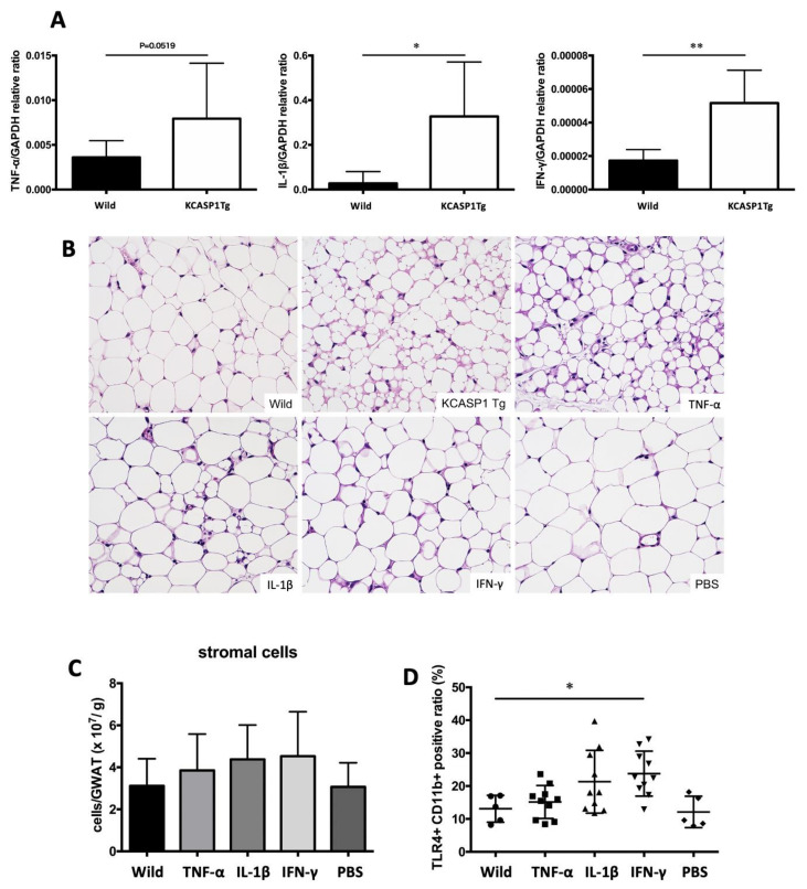
Figure 5.
Production of inflammatory cytokines in the skin and the direct effect on AT. (A) The TNFα, IL-1β and INF-γ levels were significantly increased in KCASP1Tg mouse ear skin lesions. (B) Eight-week-old BL/6 wild mice were treated by intraperitoneal injections of TNF-α, IL-1β, IFN-γ (250 µg/kg body weight/each time) or PBS three times per week for two weeks. The AT of TNF-α-treated mice showed a similar appearance to that of KCASP1Tg mice; IL-1β- and IFN-γ-administrated mice also showed a similar trend; adipocytes were large in number, small and irregularly shaped (HE, ×400). (C,D) The number of infiltrating cells in GWAT was counted, and the infiltrating cells were analyzed by flowcytometry. The number of infiltrating cells in GWAT was increased in cytokine-administrated mice, while TLR4+ CD11b+ activated monocytes were significantly increased in IFN-γ-treated mice. n = 5 per group. * p < 0.05, ** p < 0.01 versus control.

