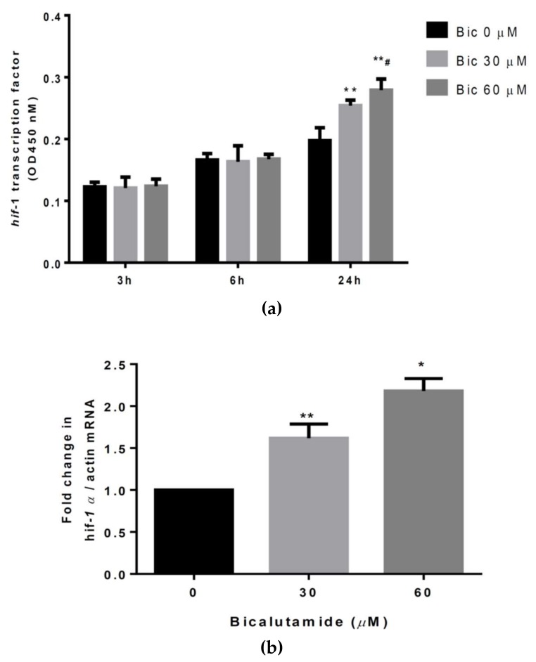
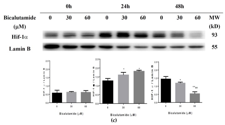
Figure 5.
(a) Hypoxia-inducible factor (HIF)-1α transcriptional activity assay. Cells were untreated or treated with bicalutamide (Bic) at doses of 30 and 60 μM for 3–24 h and hif-1α transcriptional activity was analyzed with a commercial kit (Cayman). Hif-1α transcriptional activity had only increased at 24 h after treatment with Bic. (b) Quantitative polymerase chain reaction (qPCR) for hif-1α mRNA. Rat mesangial cells (RMCs) were treated with Bic at doses of 30 or 60 μM for 24 h. Total RNA was extracted and reverse-transcribed to cDNA. hif-1α mRNA was detected according the protocol of a commercial SYBR green QPCR kit (Taigen Bioscience). Hif-1α mRNA dose-dependently increased. (c) Western blot analysis of the HIF-1α protein in RMC cells. Cells were treated with the indicated doses of Bic for 0–48 h. Nuclear proteins were subfractionated from total cell lysates and loaded onto SDS-PAGE gels. Blots were detected using antibodies of HIF-1α and lamin B. Lamin B was used as an internal control. Levels of HIF-1α protein expression were quantified by Image J software and normalized to that of lamin B. Representative images are shown and quantitative data are from three replicates. Data are expressed as the mean±standard deviation. * p < 0.05, ** p < 0.01 compared to untreated cells; # p < 0.05, ## p < 0.01 compared to 30 μM Bic-treated cells.


