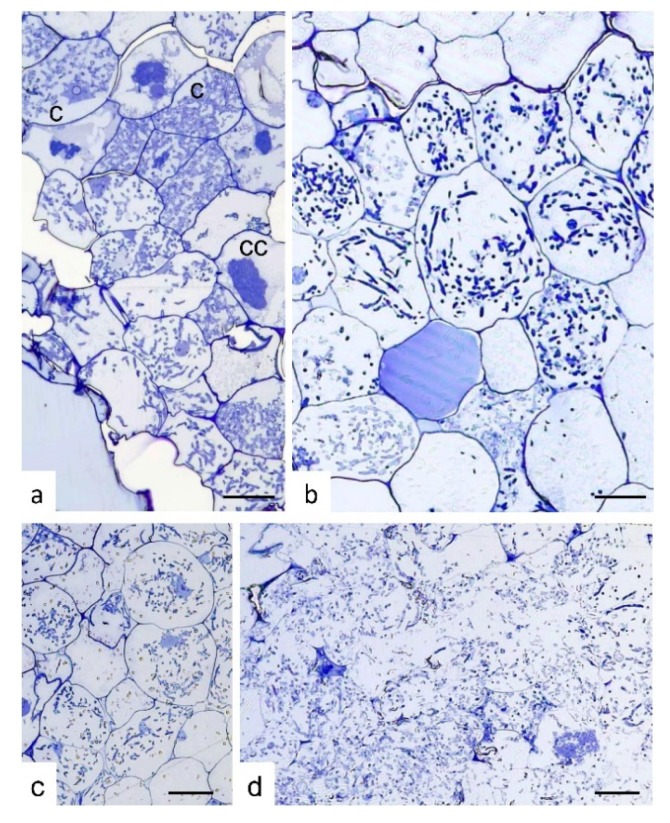
Figure 2.
Semi-thin sections of Serapias vomeracea protocorms colonized by Tulasnella calospora. (a) Stage where protocorms appeared with the typical features and color. At cellular level, typical colonization pattern with T. calospora is evident with the presence of coils at different developmental stages. c, coil; cc, collapsed coil. (b,c) Subsequent stages where protocorms are becoming brown. The fungal colonization pattern is still evident as well as host cell features. (d) Section of a dark/soft protocorm. Cell borders are not well-defined and the fungal hyphae are widespread in the tissues without a typical colonization pattern. Bars = 33, 13, 45, 25 μm for (a), (b), (c) and (d), respectively.

