Figure 4.
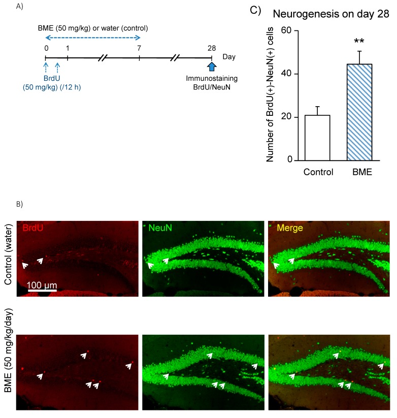
Effect of BME on neuronal differentiation of BrdU(+) cells in 5-week-old mice. (A) Experimental schedule. Animals were given either BME (50 mg/kg, p.o) or water for 7 consecutive days, and all animals received BrdU (50 mg/kg, i.p.) with a 12-h interval on the first day of the experiment and were then decapitated on day 28 post-treatment to prepare sagittal hippocampal sections. The sections were immunostained with antibodies for BrdU and NeuN. (B) Fluorescence micrographs of NeuN(+) cells (green) and BrdU(+) cells (red) in the dentate gyrus of the water- and BME-treated groups. White arrows indicate BrdU-NeuN double-positive cells. (C) Neurogenesis detected in the GCL ± SGZ of dentate gyrus on day 28 after 7-day treatment with BME. Each data column represents the mean ± SEM, calculated from four animals in each group. ** p < 0.01 compared to water-treated group.

