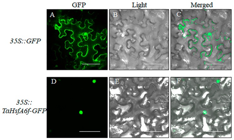
Figure 2.
Subcellular localization of TaHsfA6f protein by Nicotiana benthamiana leaf epidermal cells transient expression. 35S::GFP and 35S::TaHsfA6f-GFP plasmids were transformed into Nicotiana benthamiana leaf epidermal cells and signals were visualized with laser confocal-scanning fluorescence microscopy. From left to right, the photographs were taken in the dark-field for green fluorescence (GFP) (A,D), under bright-field for the morphology of the cell (Light) (B,E), and overlays of the GFP signals and bright-field (Merged) (C,F). The scale bars represent 40 μm.

