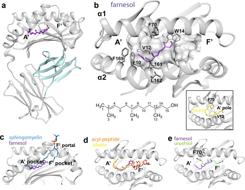
Figure 5. Crystal structure of CD1a-farnesol complexes.
a. Overview of the binary crystal structure of CD1a (grey)-farnesol (purple)/β2m(cyan). b. Molecular interactions of farnesol (purple) with the hydrophobic residues within CD1a binding cleft (grey surface). The side chains of the residues within 4 Å distance from the lipid are shown. A diagram of trans, trans farnesol with carbon numbering is shown. The A’ pole formed by V12-F70 interaction in the context of oleic acid-bound CD1a pocket (PDB ID: 4X6D) is highlighted in the inset. c-e. Superimposition of CD1a bound to farnesol and sphingomyelin (PDB ID: 4X6F, (35)) (c) lipopeptide (PDB ID: 1XZ0, (40)) (d) and urushiol (PDB ID: 5J1A, (30)) (e).

