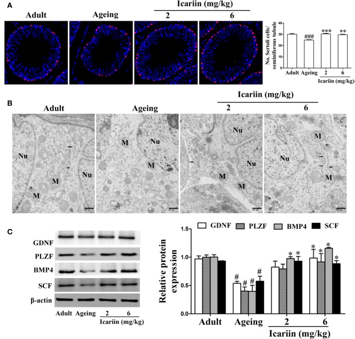
Figure 2.
Icariin protects against testicular Sertoli cell injury in aging rats. (A) The numbers of Sertoli cells in testicular tissue. Anti-SOX9 rabbit-polyclonal antibody (red) was used to detect Sertoli cell numbers, and DAPI (blue) was used for nuclear staining (original magnification, 400×). Sertoli cell numbers were measured using Image J software for more than 80 tubules per animal, with 5–8 rats in each group. (B) The ultrastructure of Sertoli cell in testicular tissues was examined using a Hitachi H-7500 transmission electron microscopy (original magnification, 20,000×; scale bar = 800 nm). These electronmicrographs are representative of three independent experiments. Nu, nuclei; M, mitochondria. Black arrowheads indicate endoplasmic reticulum. (C) The relative protein expression levels of GDNF, PLZF, BMP4, and SCF in testicular tissues were detected using western immunoblotting analysis. All values are expressed as means ± SEM of three independent experiments. #P < 0.05, ###P < 0.001 versus adult control group; *P < 0.05, **P < 0.01, ***P < 0.001 versus aging model group.

