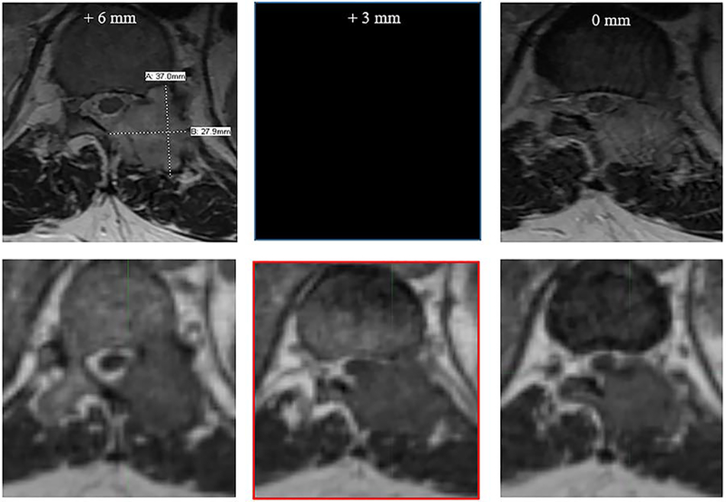
Figure 2.
Deficiencies of diagnostic MRI studies include wider than ideal slice thickness. In spinal SABR, selection of slice thickness is critical to safe and effective treatment. In the figure above, the top row represents a diagnostic axial MR image (T2 sequence), with left and rightmost images reflecting 6 mm cranio-caudal slice thickness (neighboring slices on PACS viewer). The bottom row demonstrates the 0.35T bSSFP sequence with 1.5 mm isotropic resolution. The diagnostic MRI was read as T11 nerve root compression and tumor extension into the epidural space without clear cord abutment. In the Viewray simulation, 90-degree cord abutment is readily observed (lower-central image outlined in red) between the diagnostic MRI slices.

