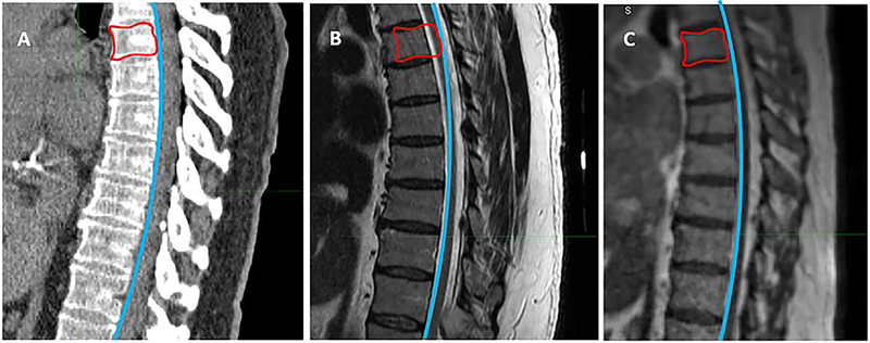
Figure 3.
MRI and CT simulation alignment. Different setup protocols for spinal diagnostic MRI can result in variable flexion, extension and rotation of the vertebral column, leading to misalignment of tumor and cord between diagnostic MRI and CT simulation. With in-house MRI-RT systems, immobilization conditions for each patient can be supervised and reproduced, making spinal curvature comparable between MR and CT scans. Image A represents a CT simulation scan, with the curve of the anterior border of the spinal canal delineated in blue and the gross tumor volume (GTV) delineated in red. Identical curves are fitted to both the diagnostic MRI (Image B) and the 0.35T MR/RT set-up scan (Image C). While the curve conforms to the spinal curvature of the MR/RT scan (Image C), subtle kyphosis in the diagnostic MRI (Image B) causes the curve to transect the spinal cord at the level of the GTV, demonstrating cord misalignment.

