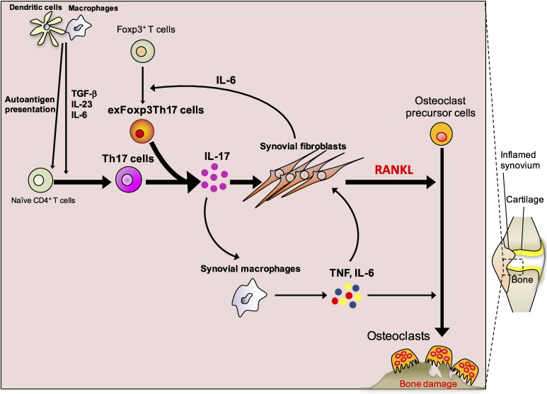
Figure 1.
(Color online) Molecular mechanisms of bone destruction in autoimmune arthritis. The autoimmune response starts with the presentation of autoantigens by antigen-presenting cells such as dendritic cells and macrophages. Naïve CD4+ T cells are activated by antigen-presenting cells and differentiate into Th17 cells, a process which is supported by various cytokines, including IL-6, IL-23 and TGF-β. Synovial fibroblast-derived IL-6 induces the conversion of Foxp3+ T cells into exFoxp3Th17 cells. Th17 cells and exFoxp3Th17 cells produce IL-17, which stimulates the expression of RANKL and inflammatory cytokines such as IL-6 in synovial fibroblasts. IL-17 also induces the production of inflammatory cytokines by synovial macrophages, including IL-6 and TNF. These inflammatory cytokines further upregulate RANKL on synovial fibroblasts and synergistically enhance RANKL-induced osteoclastogenesis.

