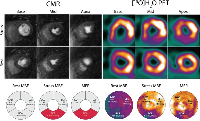
Fig. 1.
Case example of concordance between CMR and [15O]H2O PET in a 71-year-old female patient who presented with typical angina. Short-axis slices at the basal, mid, and apical levels have been selected from the PET study in order to match CMR and PET images. Both CMR and [15O]H2O PET demonstrate a perfusion defect in the inferior wall stretching from base to apex. With both techniques, the measured stress MBF and MFR in the vascular territory of the RCA are well below the ischemic thresholds. CMR = cardiac magnetic resonance imaging; LAD = left anterior descending artery; LCx = left circumflex artery; MBF = myocardial blood flow; MFR = myocardial flow reserve; PET = positron emission tomography; RCA = right coronary artery

