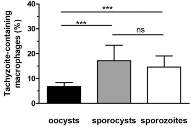
Figure 4.

Development of tachyzoites in RAW macrophages challenged with oocysts, free sporocysts, or sporozoites. Following parasite-macrophage incubation for 24 h, the percentage of tachyzoite-containing macrophages was determined by immunofluorescence assay as described in materials and methods. Mean ± standard deviation of 4 independent experiments. One-way ANOVA and Tukey's post-test, ***p < 0.001.
